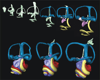
University of Iowa researchers and their colleagues have developed imaging techniques that may allow researchers and physicians to better understand normal inner ear development as well as inner ear diseases.

Graduate student Benjamin Kopecky and Bernd Fritzsch, professor and departmental executive officer in the UI Department of Biology, published their results in the March issue of the journal Developmental Dynamics.
Using an improved 3D analysis of mouse ears during normal embryonic development, the researchers showed:
- New insights into ear development.
- A technique permitting the first full quantification of volumetric and linear aspects of ear growth.
- Data providing the framework for future analysis of mutant conditions currently under-appreciated using only two-dimensional imaging.
Regarding the benefits of their 3D imaging technique, the researchers say current 3D reconstructed images are limited by a paint-filling process that leaves images distorted. Also, the segmentation process used to create images takes significant time. Meanwhile, current microcomputer tomography (CT) and high-resolution magnetic resonance imaging yield low resolution—sufficient for the human ear, but not for smaller samples.
Physicians could use the new process to provide faster, better images that may benefit patients suffering from inner ear diseases by correlating human ear defects with defects in mice that have fully characterized mutations. The process works by using a thin-sheet laser imaging microscope (sTSLIM) to compile high-resolution images of the ear through nondestructive computerized optical sections.
In addition to Kopecky and Fritzsch, coauthors of the paper include Shane Johnson, Heather Schmitz, and Peter Santi, all of the University of Minnesota Department of Otolaryngology.
The UI Department of Biology is part of the UI College of Liberal Arts and Sciences.
The research was sponsored in part by the National Institutes of Health, National Institute on Deafness and Other Communication Disorders.