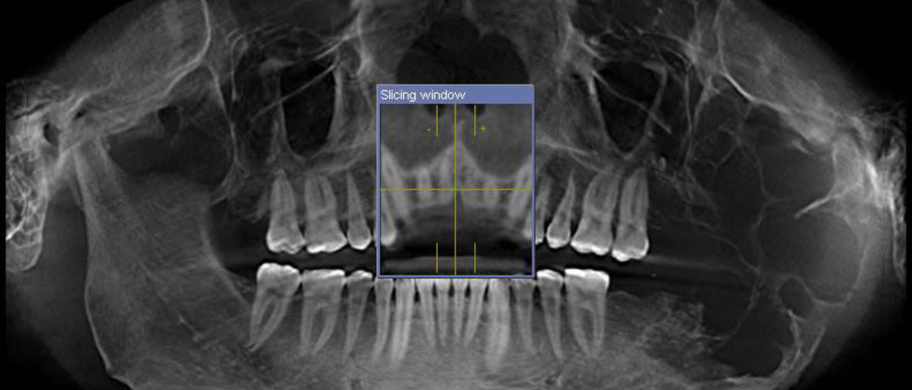
A pair of faculty at the University of Iowa this month will publish a new edition of Tumors and Cysts of the Jaws, considered the go-to guide for developments in the field of jawbone diseases and diagnosis. The authors, Robert Robinson, pathology professor in the Carver College of Medicine, and Steven Vincent, professor and head of oral pathology, radiology and medicine in the College of Dentistry, discuss their collaboration.
You specialize in diagnosing tumors and cysts in the human jaw. What are some examples and how prevalent are they?
Robinson and Vincent: The prevalence of jaw tumors is on-par with similar conditions involving other bones of the skeleton. Significantly, a number of tumors and cysts that occur in the upper and lower jawbones are not found anywhere else and have unique clinical and microscopic features. Some behave aggressively requiring additional treatment to help prevent recurrences; others can be treated in a very simple manner. The pathologist must be familiar with the sometimes subtle differences between these aggressive and non-aggressive tumors when communicating with the surgeon.
How long did it take to do this volume? How did you first both get involved?
Robinson and Vincent: We have a long collaborative history, including numerous continuing education programs on this subject given at the United States and Canadian Academy of Pathology and the American Academy of Oral and Maxillofacial Pathology annual meetings. Dr. Robinson was contacted by Dr. Steven G. Silverberg, the editor-in-chief of the Armed Forces Institute of Pathology, American Registry of Pathology, regarding this volume. It was only natural that we should work on this publication together.
What is new in this edition and what does it mean for the field?
Robinson and Vincent: The art and science of pathology is a very dynamic area of health care. New types of tumors and cysts are defined frequently, which is very important in terms of treatment and prognosis. This volume includes the latest information regarding this unique area of pathology since the volume’s last publication over 12 years ago. Additionally, volume IV contains numerous high definition microscopic images, radiographs and CT scans, which are sometimes necessary for a specific diagnosis.
How has the field of jaw pathology diagnostics changed since you entered it?
Robinson and Vincent: Pathology now relies more on special staining methods and molecular biology techniques, which were not available when we began our careers. Volume IV reflects this -- for the first time it contains contemporary laboratory procedures that tell us more about the types and behaviors of the tumors and cysts we diagnosis, and often gives us better information about the expected behavior, which is of greatest importance for patient management.
