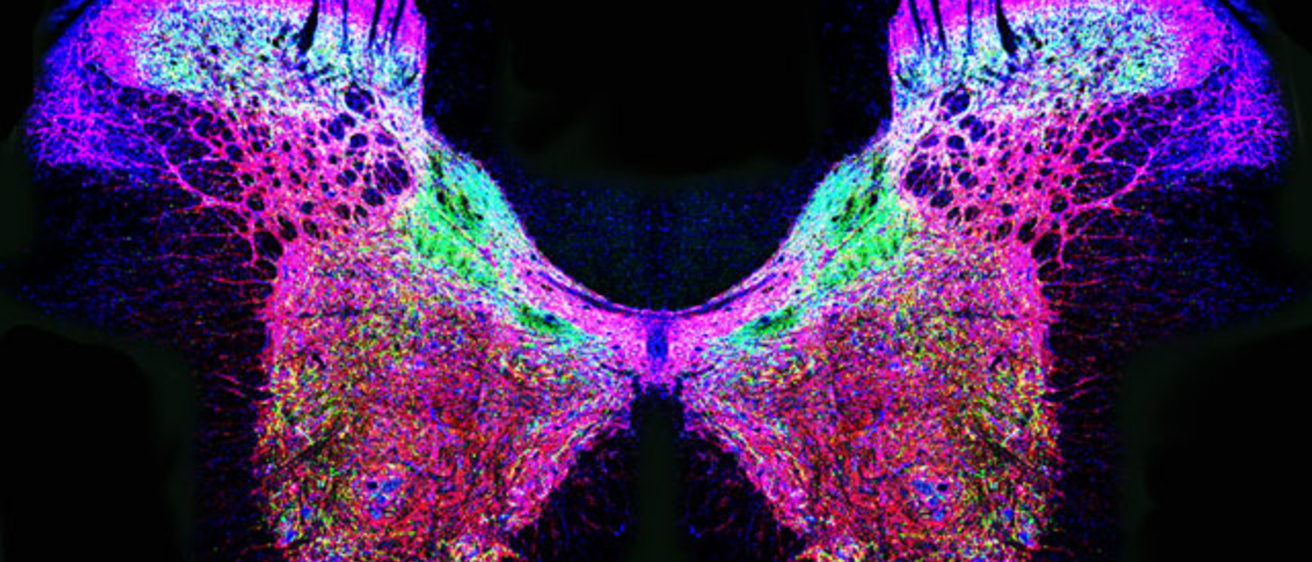
It may look like an exotic butterfly, but this image actually shows the location in the spinal cord of three enzymes that are critical for breathing and heart function.
The image, which was created by University of Iowa neuroscientist Li-Hsien Lin, Ph.D., was one of 10 winning entries of Bio-Art, the Federation of American Societies of Experimental Biology's (FASEB) first-ever biomedical image competition.
The winning images were unveiled on FASEB's website, displayed during the organization's 2012 Centennial Reception on May 16 at the United States Capitol Complex in Washington D.C., and will be featured on the National Institutes of Health (NIH) website and main campus in Bethesda, Md.
Lin is an associate research scientist in the Neurobiology and Circulatory Control Laboratory run by UI professor of neurology William Talman, M.D. She also is a member of the American Physiological Society.
She used confocal microscopy to create the image of a rat spinal cord showing the distribution of three types of glutamate and nitric oxide synthesizing enzymes. Both glutamate and nitric oxide play an important role in transmitting cardiovascular and respiratory signals between the brain, heart, and lung. Understanding the action and interaction of glutamate and nitric oxide in the nervous system could lead to better treatments for cardiovascular diseases such as hypertension and heart failure. The work is supported by NIH funding from the National Heart, Lung and Blood Institute.
The Bio-Art competition aimed to recognize the most captivating representations of cutting edge biomedical research. To view all the winning images, visit www.faseb.org/Scientific-Image-Competition/Winners.aspx.
