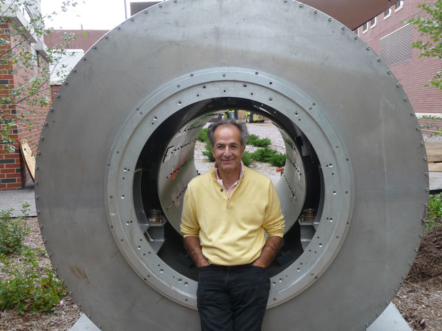
This fall, the University of Iowa will get a powerful imaging device, allowing researchers to scan anywhere in the human body without so much as a prick or a poke. The UI community and the public can get a sneak preview of the new magnetic instrument at a symposium this Thursday, Sept. 12.

The arrival of the 7 Tesla Scanner in November will make the UI one of only about 20 research institutes in the United States—and only about 30 worldwide—with the instrument. The giant magnet's arrival should be something to behold: Weighing 36 tons, or about 6,000 gallons of paint, it will travel overseas by barge from the United Kingdom, where it was built, be lifted by crane and deposited on the first floor of the Pappajohn Biomedical Discovery Building, currently under construction.
“The 7T scanner will permit us to remain at the forefront of magnetic resonance imaging (MRI) research and to conduct studies that simply were not previously possible,” says Vince Magnotta, associate professor in radiology and director of the UI’s Magnetic Resonance Imaging facility, which won a $8 million grant from the National Institutes of Health (NIH) to purchase the magnet.
The symposium will be held from 8:15 a.m. to 12:45 p.m. in the Urmila Sahai Seminar Room, Medical Education and Biomedical Research Facility, on the UI medical campus. The keynote speaker will be Kamil Urgurbil, a member of the Brain Research through Advancing Innovative Neurotechnologies Committee, a NIH-led initiative to increase our understanding of the human brain. There also will be talks by UI faculty in dentistry, orthopaedics and rehabilitation, psychiatry, psychology, and radiology on how the scanner can be used.
The 7T scanner will open research opportunities, such as allowing scientists to study metabolism and microstructures in the body only possible with ultra-high field devices. Moreover, the scanner will yield clearer, higher-resolution images of the brain, enhancing researchers’ ability to study the brain’s function and connectivity, according to psychiatry professor Nancy Andreasen, who has worked with magnetic imaging equipment for 30 years.
The scanner will not just be for the medical community. Investigators in the Tippie College of Business, for example, plan to use it for work in the emerging field of neuroeconomics, or how the brain engages in everyday decision making.
The instrument, which will be in the Iowa Institute for Biomedical Imaging, will add to the university’s legacy in imaging research. UI researchers conducted the first magnetic resonance imaging study to identify the presence of brain abnormalities in patients with schizophrenia in the early 1980s, studies led by Andreasen.
Since then, thousands of magnetic resonance studies have been conducted and scanners have become popular research tools used by investigators across a variety of fields. Prior to MRI, researchers were limited to viewing the body’s structures and organs through invasive surgeries and often only after a person had died.
Individuals with disabilities are encouraged to attend all UI-sponsored events. If you are a person with a disability who requires a reasonable accommodation in order to attend this symposium, call 319-384-5232 in advance.